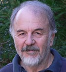
Harry Noller

Professor Emeritus of MCD Biology
Robert L. Sinsheimer Prof. of Molecular Biology
Director, Center for Molecular Biology of RNA
A.B., University of California, Berkeley
Ph.D., University of Oregon
1965-66 Natl Inst of Health Postdoctoral Fellow,
MRC Laboratory of Molecular Biology, Cambridge
1966-68 Natl Inst of Health Postdoctoral Fellow, Inst of Molecular Biology, Univ of Geneva, Switzerland
Ribosome Structure and Function
Ribosomes are complex molecular machines that are responsible for carrying out protein synthesis – translation of the genetic code. Their structures are highly conserved, and fundamentally similar in all organisms. A major question is, why do ribosomes contain such large amounts of RNA (50-60% of their mass)? It has gradually become clear that ribosomal RNA itself is centrally involved in translation, and may be a relic of the RNA World, a period of early molecular evolution when it is proposed that RNA carried out the genomic and catalytic roles of DNA and protein.
The Noller laboratory studies ribosome structure and function using a wide range of approaches, including X-ray crystallography, chemical probing methods, molecular genetics, comparative sequence analysis and fluorescence resonance energy transfer (FRET), including the use of single-molecule methods. The ultimate goal of these studies is to understand how the ribosome works at the molecular level: what are the moving parts of the machine, and how do they move in three dimensions to enable translation?
X-ray Crystal Structure of the Ribosome
We originally solved the structure of the whole 70S ribosome containing mRNA and three tRNAs bound in the A, P and E sites at a resolution of 5.5 Angstroms (Yusupov et al., 2001). This enabled us to describe the positions and interactions of the ribosome's functional ligands. Next, we solved the all-atom structures of different ribosome functional complexes, including release factors RF1, RF2 and RF3, and elongation factor EF-G (Laurberg et al., 2008; Korostelev et al., 2008, 2010; Zhou et al., 2011, 2012, 2013, 2014). These structures have led to insights into the molecular mechanisms of translation termination and translocation of mRNA and tRNA. Our long-term goal is to re-create a three-dimensional movie of the ribosome carrying out protein synthesis at the atomic level, by solving structures of the ribosome trapped in many different intermediate states of translation. This challenging project will require close coordination between ribosome biochemistry and crystallography.
Movement Inside the Translational Engine
On the basis of chemical footprinting results, we realized that the tRNAs move on the ribosome during the translocation step of protein synthesis in two steps: first they move at their acceptor CCA ends on the large ribosomal subunit, and then they move at their anticodon ends on the small subunit (Moazed & Noller, 1989). Although the first step can proceed spontaneously (Cornish et al., 2008), the second step is dependent on elongation factor EF-G. Recently, we have solved the crystal structures of ribosome complexes trapped in intermediate states of translocation. These structures show that rotation of the head domain of the small ribosomal subunit is involved in movement of the mRNA and anticodon stem-loop ends of the tRNAs in the small subunit (Zhou et al., 2013). This rotational movement of the 30S subunit and its head domain as well as the movement of the L1 stalk of 23S rRNA during translocation have also been observed directly using FRET (Ermolenko & Noller, 2011; Guo & Noller, 2012; Cornish et al., 2009). In collaboration with the Tinoco and Bustamante groups at UCB, much has also been learned about the movements and forces within the ribosome using single-molecule optical tweezers approaches (Liu et al., 2014; Qu et al., 2012; Wen et al., 2008). Elements of the tRNA binding sites in both subunits are dynamic; we have recently discovered that ribosomal RNA elements of the P site reach toward the A site to contact the tRNA and escort it into the P site during EF-G-catalyzed translocation (Zhou et al., 2014). Finally, the structure of tRNA itself flexes dramatically as it moves through the ribosome during translocation. These findings all point to the importance of RNA structural dynamics for protein synthesis, consistent with the proposed origins of the ribosome from an RNA world (Noller, 2011).
Some Unanswered Questions
Many mysteries remain concerning the molecular mechanisms underlying protein synthesis, particularly the translocation process. Why does the first step of translocation proceed spontaneously, but not the second step? Rotation of the small-subunit head domain moves the mRNA and tRNAs forward on the small subunit; how does the head rotate back without pulling the tRNA and mRNA back with it? How does elongation factor EF-G overcome the energy barrier to the second step? What does GTP hydrolysis by EF-G accomplish? Is interdomain movement of EF-G directly responsible for movement of mRNA and tRNA from the A site to the P site? How does elongation factor EF-Tu overcome the steep kinetic barrier to accommodation of aminoacyl-tRNA into the large subunit A site during the step of aminoacyl-tRNA selection? Does this involve structural rearrangements in the large subunit? Finally, how are all of the different structural movements inside the ribosome coordinated and controlled during protein synthesis?
For a full list of publications, please follow this link.
Colussi, T.M, Costantino, D.A., Zhu, J., Donohue, J.P., Korostelev, A.A., Jaafar, Z., Plank, T.M., Noller, H.F., and Kieft, J.S. (2015) A structured eukaryotic IRES RNA can initiate translation in bacteria. Nature 519:110-113.
Zhou, J., Lancaster, L., Donohue, J.P. and Noller, H.F. 2014. How the ribosome hands the A-site tRNA to the P site during EF-G-catalyzed translocation. Science 345:1188-1191.
Mohan, S., Donohue, J.P and Noller, H.F. 2014. Molecular mechanics of 30S subunit head rotation. Proc Natl Acad Sci U S A 111:13225-13230.
Liu, T., Kaplan, A., Wickersham, C.E., Wen, J.-D., Alexander, L., Lancaster, L., Fredrick, K., Noller, H.F., Tinoco, I. Jr. and Bustamante, C. 2014. Direct Measurement of the Mechanical Work During Translocation by the Ribosome. eLife 3:e03406.
Ramrath, D.J.F., Lancaster, L., Sprink, T., Mielke, T., Loerke, J., Noller, H.F. and Spahn, C.M.T. 2013. Visualization of two tRNAs trapped in transit during EF-G-mediated translocation. Proc Natl Acad Sci U S A 110:20964-20969.
Noller, H.F. 2013. How does the ribosome sense a cognate tRNA? J. Mol. Biol. 425:3776-3777.
Noller, H.F. 2013. By Ribosome Possessed. J. Biol. Chem. 288:24872-24885.
Zhou, J., Lancaster, L., Donohue, J.P. and Noller, H.F. 2013. Crystal Structures of EF-G-Ribosome Complexes Trapped in Intermediate States of Translocation. Science 340:1236086
Guo, Z. and Noller, H.F. 2012. Rotation of the head of the 30S ribosomal subunit during mRNA translocation. Proc Natl Acad Sci U S A 109:20391-20394.
Zhou J, Korostelev A, Lancaster L and Noller HF. 2012. Crystal structures of 70S ribosomes bound to release factors RF1, RF2 and RF3. Curr Opin Struct Biol. 2012 22:733-742
Qu, X., Lancaster, L., Noller, H.F., Bustamante, C. and Tinoco, I., Jr. 2012. Ribosomal protein S1 unwinds double-stranded RNA in multiple steps. Proc Natl Acad Sci U S A 109:14458-14463.
Zhou, J., Lancaster, L., Trakhanov, S. and Noller, H.F. 2011. Crystal Structure of Release Factor RF3 Trapped in the GTP state on a rotated conformation of the ribosome. RNA 18:230-240.
Qu, X., Wen, J.D., Lancaster, L., Noller, H.F., Bustamante, C., and Tinoco, I., Jr. 2011. The ribosome uses two active mechanisms to unwind messenger RNA during translation. Nature 475: 118-121.
Zhu, J., Korostelev, A., Costantino, D.A., Donohue, J.P., Noller, H.F. and Kieft, J.S. 2011. Crystal structures of complexes containing domains from two viral internal ribosome entry site (IRES) RNAs bound to the 70S ribosome. Proc Natl Acad Sci USA 108:1839-1844.
Ermolenko, D.N. and Noller, H.F. 2011. mRNA Translocation Occurs During the Second Step of Ribosomal Intersubunit Rotation. Nat Struct Mol Biol 18:457-462.
Noller, H.F. 2011. Evolution of Protein Synthesis from an RNA World. In: RNA Worlds, 4th Ed. (eds. J.F. Atkins, R.F. Gesteland, T.R. Cech) Cold Spring Harbor Laboratory Press, Cold Spring Harbor, New York. pp.141-154.
Korostelev, A., Zhu, J., Asahara, H. and Noller, H.F. 2010. Recognition of the amber UAG stop codon by release factor RF1. EMBO J 29:2577-85.
Korostelev, A., Laurberg, M., and Noller, H.F. 2009. Multistart simulated annealing refinement of the crystal structure of the 70S ribosome. Proc Natl Acad Sci USA 106:18195-18200.
Cornish, P.V., Ermolenko, D.N., Staple, D.W., Hoang, L., Hickerson, R., Noller, H.F. and Ha, T. 2009. Following Movement of the L1 Stalk Between Three Functional States in Single Ribosomes. Proc Natl Acad Sci USA 106:2571-2576.
Korostelev, A., Asahara, H., Lancaster, L., Laurberg, M. Hirschi, A., Zhu, J., Trakhanov, S., Scott, W. and Noller, H.F. 2008. Crystal Structure of a Translation Termination Complex Formed with Release Factor RF2. Proc Natl Acad Sci USA 105:19684-19689.
Korostelev, A., Ermolenko, D.N. and Noller, H.F. 2008. Structural Dynamics of the Ribosome. Curr Opin Chem Biol 12:674-83
Lancaster, L., Lambert, N.J., Maklan, E.J., Horan, L.H and Noller, H.F. 2008. The sarcin-ricin loop of 23S rRNA is essential for assembly of the functional core of the 50S ribosomal subunit. RNA 14:1-14.
Martick, M., Horan, L., Noller, H., and Scott, W.G. 2008. A discontinuous hammerhead ribozyme embedded in a mammalian messenger RNA. Nature 454:899-903.
Laurberg, M., Asahara, H., Korostelev, A., Zhu, J., Trakhanov, S. and Noller, H.F. 2008. Structural basis for translation termination on the 70S ribosome. Nature 454:852-857.
Wen, J.D., Lancaster, L., Hodges, C., Zeri, A.C., Yoshimura, S.H., Noller, H.F., Bustamante, C., and Tinoco, I. 2008. Following translation by single ribosomes one codon at a time. Nature 452:598-603.
Cornish, P.V., Ermolenko, D.N., Noller, H.F., and Ha, T. 2008. Spontaneous intersubunit rotation in single ribosomes. Mol Cell 30:578-88
